
8 - Inspection and test techniques
8.4 - Acoustic microscopy, electron microscopy
Acoustic microscopy
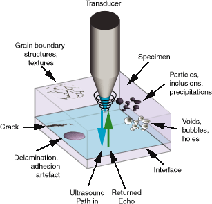
Scanning Acoustic Microscopy (SAM) is a non-destructive method for revealing cracks, breaks and voids that cannot be shown by X-ray microscopy. Ultrasonic waves are focusable mechanical waves with a frequency above 20 kilohertz. Materials have a property which characterises their capability of decrease the velocity of sound in them. This is acoustic impedance:where is the acoustic impedance, is the density of the material, and is the velocity of the wave in that material. From the interfaces of materials with different acoustic impedances, the ultrasonic wave is either absorbed, scattered or reflected. It is highly sensitive to the elastic properties of the materials it travels through, and is especially sensitive to air gaps. The wave firstly reflected gets firstly to the transceiver. The time between detected waves is determined by the distance between the interfaces. This makes the acoustic microscopy the preferred method for finding delaminations, cracks, voids and porosity. From the time delay, the thickness of the structures can also be determined.
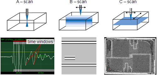
There are three main modes of imaging in acoustic microscopy. At A-scan, the transducer is above a single point where it transmits and receives the signal, the waves reflected from that point. With the software, we can select time windows that are time gates with optional lengths to get the signal from the desired depth. At B-scan, the transducer is scanning by a line of X-Z or X-Y direction. A vertical cross section is displayed of the specimen. It is a line of A-scans. At C-scan, the scanning is in both X and Y directions. A horizontal cross-section image is made of the surface or an inner layer of the specimen. Waves reflected from the same depth can be selected with time gates.
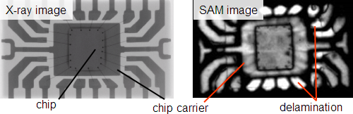
Acoustic microscopes are most commonly used in failure analysis, but there are in-line SAM machines either. The following failures can be inspected: delamination, die-attach, voids, cracks, and pseudocoloring of digital images helps to highlight them.
Electron microscopy
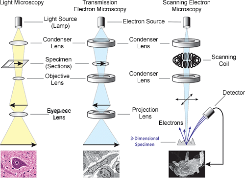
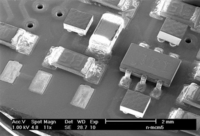
Out of all imaging methods, the highest magnification and resolution can be reached with electron microscopy (EM), because the wavelength of electron-ray is much less than the wavelength of light or X-ray. Electrons are coming from the electron gun. They are focused or deflected - to scan - by magnetic field produced by magnetic coils. Allegorically, these coils function as magnetic electron lenses. To avoid the scattering of electrons from gas particles, the whole process takes place in vacuum. The electron detectors are situated just above the sample. The main types of electron microscopes are transmission electron microscope (TEM), and scanning electron microscope (SEM). For TEM, the specimen should be a thin, film-like sheet. The throughpassing electrons form the image, according to the atomic or molecular structure of that specimen. For SEM, the surface of the specimen is scanned and the backscattered electrons or the sample current is measured for imaging. The sample current is of the electrons which fall to the sample. With electron microscopy, only solid, electronically conductive specimen or samples coated with thin conductive layer can be examined. This layer is of gold or carbon and its task is to avoid electrostatic charging.
Electron-matter interaction, imaging
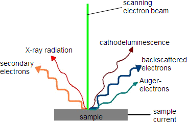
The electron beam excites the material. From the electron-matter interaction, secondary electrons, backscattered electrons and the sample current is produced and used for imaging. X-ray radiation and Auger-electrons are used for spectroscopy - identification of substance. Secondary electrons are produced when the electron inelastically collide the atom of the sample, and knocks out valence electrons. Backscattered electrons are electrons which are elastically scattered back from the surface. After emitting a secondary electron, a high orbital energy electron falls to this empty, lower energy orbit. The energy difference is radiated out of the atom in the form of characteristic X-ray radiation. Auger-electrons are made almost by the same way: here, the energy difference is passed to another electron and it departs. Characteristic X-ray radiation: after emitting a secondary electron, a high orbital energy electron falls to this empty, lower energy orbit; the energy difference is radiated out of the atom. An important thing in electron microscopy is the setting of accelerator voltage. Accelerator voltage produces a magnetic field, which increases the velocity of electrons. If it is higher, the image is shinier, but the resolution and the depth of field is worse.
The electron beam excites the material. From the electron-matter interaction, secondary electrons, backscattered electrons and the sample current is produced and used for imaging. X-ray radiation and Auger-electrons are used for spectroscopy - identification of substance. Secondary electrons are produced when the electron inelastically collide the atom of the sample, and knocks out valence electrons. Backscattered electrons are electrons which are elastically scattered back from the surface. After emitting a secondary electron, a high orbital energy electron falls to this empty, lower energy orbit. The energy difference is radiated out of the atom in the form of characteristic X-ray radiation. Auger-electrons are made almost by the same way: here, the energy difference is passed to another electron and it departs. Characteristic X-ray radiation: after emitting a secondary electron, a high orbital energy electron falls to this empty, lower energy orbit; the energy difference is radiated out of the atom. An important thing in electron microscopy is the setting of accelerator voltage. Accelerator voltage produces a magnetic field, which increases the velocity of electrons. If it is higher, the image is shinier, but the resolution and the depth of field is worse.
An electron microscopy examining station composes of an electron microscope, a vacuum system and a computer. With scanning electron microscopes, meaningful images can be made of electronic components and materials. Images of biological samples are also well-known.
| Previous | Next |