
7 - Failure analysis
7.1 - Microscopy techniques
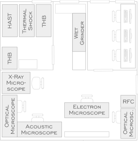
Floor-plan of the failure analysis laboratory
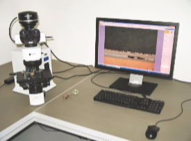
Optical microscopy
The Olympus BX-51 stereo microscope is connected to a computer through a high resolution CCD camera. Besides the convenient visual inspection, it is also possible to measure distances and angles. Saving images for further processing and printing is also supported.
X-Ray inspection
The DAGE XiDAT XD6600 X-Ray microscope is used for inspecting failures e.g. in solder joints, such as open joints, solder bridging, voids etc.
Acoustic microscopy
Ultrasound wave generated by the transducer travels to the specimen through a medium called couplant (usually deionized water). The wave then travels through the specimen's material at the material's velocity, with a portion of it being reflected back everytime it hits an interface within the material. In the pulse echo method, the same transducer is used as sender and receiver of the sound waves. The echoes received by the transducer are converted to voltages, amplified, digitized, and presented as an image. (See: A-, B-, and C - Scans)
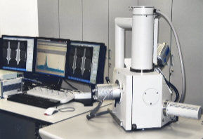
Scanning Electron Microscope
The FEI Inspect S50 Scanning Electron Microscope is used for general high magnification imaging and measurements, and for material composition analysis of micro-scale volumes using energy dispersive X-Ray spectroscopy.
| Previous | Next |