Picture

6 - Application of sensors

6.4 - Biomedical sensors

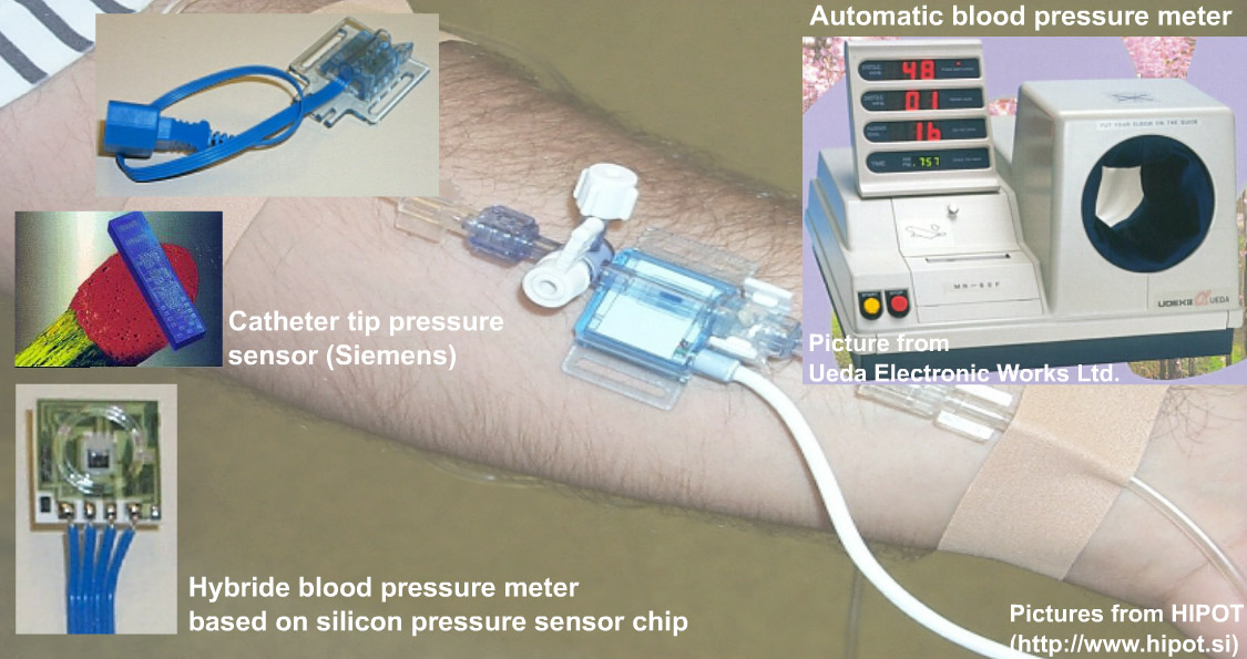
Blood pressure meter
Tonometer
An important application field of miniature silicon based mechanical sensors may be the intraocular pressure measurement. The internal pressure (p) in a sphere can simply calculated be by measuring the necessary force (F) that can flatten a certain area (A) of the sphere: p=F/A. The sensor consists of a silicon substrate containing four meander-shaped diffused resistor arrays, having a number of resistor elements with meander contacts at the resistor nodes. The sensor is covered by a Mylar (DuPont) foil, which is coated with a thin gold layer at the side that faces the sensor surface. The foil is separated from the sensor by polyimide spacer. When the applanation sensor is pressed against the eye globe, the Mylar foil with its gold film contacts a certain area as a function of the applied force; thus, shortcircuiting the meander resistor arrays within that area. Measuring the resulted resistance values that reflect the size and eccentricity of the flattened area, its diameter and distance from the nearest edge can be calculated. (A.P. Bergveld)
Radiology
X-Ray diagnostics
X-ray radiation is the oldest and even nowadays most widely used tool of noninvasive medical imaging in radiological diagnostics. In the classical X-ray diagnostics, visible images are produced either by means of an X-ray-sensitive film or by displaying on a fluorescent screen. In the latter case, X-ray image amplifiers enable the direct image display. Incident X-rays are impinging onto the surface of a fluorescent (generally CsI) screen from which secondary, visible photons are emitted. These photons excite photoelectrons from the photo-cathode of a vacuum tube which, by means of an appropriate electron optics, can be used to get a real-time visible image on a display screen located on the opposite side of the same vacuum tube. Combining the X-ray amplifier with image dividers and video cameras, not only the real-time monitoring but also image and motion storage becomes available.
Computer tomography
X-ray Computer Tomography (CT) was the greatest invention in the history of X-ray radiology. Images are made not parallel to the axis of the human body, but perpendicularly, enabling cross-sectional view preparation. The basic idea, independently from the practical appliance realizations, is that the collimated X-ray bundle sweeps the body transversally and the penetrated radiation intensity profile versus angle is measured by the detector array located on the opposite side of the body The X-ray source moves around the object making intensity profiles in a number of positions which are then processed by the computer.
Blood oxygen sensors
A new type transcutaneous pO2 sensor structure is presented here, which is based on multilayer ceramic technology. The electrodes are made by thick film technology using small amounts of precious metals. The heating element is an integrated thick film resistor and a pn-junction is used for temperature sensing. The device works on 43.5 oC temperature. The ceramic body was molded into a plastic package. The PTFE membrane with a thickness of 10 um was fastened with a mounting ring to the surface of the sensor. A conventional KCl solution with a buffering agent was used as electrolyte. A thin disc-shaped cellophane foil was placed between the electrodes and the membrane as a spacer.
Glucose sensors


Previous Next